 |
| |
 |
Uveodermatologic Syndrome |
|
|---|---|---|
| ||||||
|
Uveodermatologic Syndrome in A Two Year Old Female
Akita
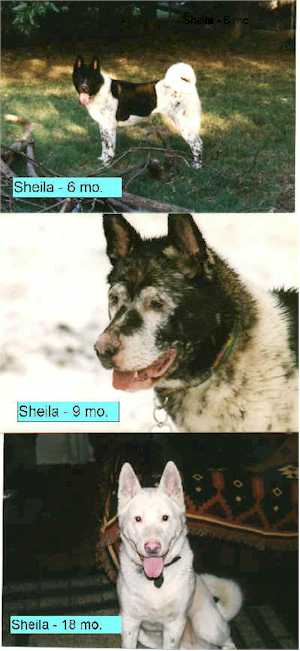
History Name: "Sheila" Date of Birth: 10/01/96 Sex: Spayed female Color: Black and white Weight: 91 lbs. (41.4 kg) At 9 months of age, Sheila’s black pigmentation began fading to the point she appears now appears albinotic (Figure 1). The owners noted that her vision problems began 5/28/98 and consulted with the referring veterinarian. They noted she had trouble distinguishing large objects and differentiating dogs, people, etc. She was worse in dim light and her eyes seemed sensitive to light, becoming "red and bloodshot" in bright light. After initial phone consultation by the referring veterinarian with the ophthalmologist, the dog was begun on a regimen of oral prednisone and azathioprine (Imuran). The initial dose of prednisone was 1.1 mg./kg. twice daily until signs abated (7-10 days); then once daily for 2 weeks; then every other day for 2 weeks. Azathioprine was initiated at one 50-mg. tablet (approximately 1.1 mg./kg. once daily for 1 week, then every other day thereafter on a maintenance dose. In the event of exacerbation of the inflammation, prednisone dosing was resumed for a short term as needed to control the attack. Upon presentation to the ophthalmologist (10/12/98) the problem had been controlled on azathioprine alone for approximately 8 months and no further deterioration of vision had occurred. In addition, laboratory assessments (CBC and chemistry panels) had been performed periodically by the referring veterinarian and were unremarkable. Ophthalmologic Examination The Schirmer tear test results were 25 and 20 mm/minute in the right and left eyes respectively. Applanation tonometry with a Tonopen-XL (Mentor) revealed intraocular pressures of 10 and 7 mmHg in the right and left eyes respectively. Generalized depigmentation of the skin, including the lips, nose, lids and ocular adnexae was present. The corneas had superficial central ring-shaped opacities and the lenses were normal. No aqueous flare was noted, and the anterior chambers were of normal depth. Indirect ophthalmoscopic examination revealed depigmentation of the retinal pigmented epithelium and choroid in the central fundus while the overlying retina appeared normal. Direct and indirect pupillary light reflexes (PLR’s) were normal, and the dazzle and menace responses were present. The patient was able to negotiate around obstacles in normal room lighting. Treatment The treatment with azathioprine was continued at one 50-mg. tablet every other day with instructions for continued monitoring of CBC and serum chemistries by the referring veterinarian at 2-3 month intervals. Topical application of 1% prednisolone sodium phosphate ophthalmic solution twice daily was added to the treatment regimen to better control a mild subclinical iridocyclitis. Discussion This case is interesting in that it is a textbook picture of uveodermatolgic syndrome. Indeed, Herrera and Duchene recently presented a report of a similar case in a dachshund and noted that 80.5% of the cases presented in the literature affected Japanese akitas.1 The disease is similar to Vogt-Koyanagi-Harada (VKH) syndrome in man which is characterized by a bilateral granulaomatous uveitis, diffuse vitiligo and poliosis, and meningitis. Tinitis or other auditory disturbances often characterize the meningitis in man. Since such auditory disturbances may not be readily obvious in dogs, the term uveodermatologic syndrome (UDS) has been used in veterinary medicine in place of VKH. The disease is generally thought to be an autoimmune disease against melanocytes, but the exact etiology and pathologic mechanisms remain unclear. The high incidence in the Akita is consistent with a disease influenced by genetic or hereditary factors. It is interesting in the case reported here that the irides appear hyperpigmented when other pigmented tissues are depigmented. It is possible that the iridal melanocytes are somehow antigenically distinct or are not affected to the same degree as other uveal dermal melanocytes. It is also possible that the irides were initially depigmented and have returned to a more normal and more heavily pigmented state as was suggested by Herrera and Duchene in their case.1 The low intraocular pressures observed at the time of ophthalmic examination indicates that a continuing subclinical iridocyclitis is present and emphasizes the need for long term immune suppressive therapy in these cases. Periodic analyses of CBC and serum chemistries are indicated since azathioprine can be hepatotoxic and can also cause bone marrow suppression. While UDS can cause blindness in association with secondary retinal detachment, cataracts, and glaucoma, determined immunosuppressive therapy and care can arrest the disease and maintain vision. Premature attempts to reduce or discontinue therapy are ill advised and often result in blindness. References 1. Herrera, HD and Duchene, AG. Uveodermatologic syndrome (Vogt-Koyanagi-Harada-like syndrome) with generalized depigmentation in a dachshund. Veterinary Ophthalmology 1998; 1(1): 47-51.
| |||||||||||
|
|