We are frequently asked the following questions
regarding cataracts in animals. The answers are intended as general responses to increase
your understanding. Please feel free to ask any additional questions you may have.
WHAT ARE CATARACTS?
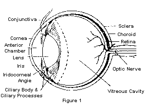
A cataract is defined as any opacity (or opacities) of
the lens of the eye (see Figure 1). Such opacities may be quite small and interfere little
with vision, or they may involve the entire lens causing blindness. Cataracts occur
because something interferes with the normal function of lens fibers causing them to
degenerate. Causes include inflammatory diseases, hereditary factors, aging changes,
toxicities, and metabolic diseases such as diabetes mellitus.
DO ALL ANIMALS DEVELOP CATARACTS WITH AGE?
In general, no! In fact most animals should live their
entire lives without developing cataracts. The lens does become thicker with age and thus
appears grayer causing many people to mistake this change for a cataract. This normal
aging process is called nuclear or lenticular sclerosis and does not impair vision other
than making focusing on close objects more difficult. However some animals do develop
cataracts and certain breeds of animals are afflicted with hereditary cataracts so that a
significant percentage of the population may develop cataracts. This is especially true in
dogs. Miniature schnauzers, cocker spaniels, poodles, Labrador retrievers and golden
retrievers are just a few of the breeds which may be affected.
HOW DO YOU TREAT CATARACTS?
There is no effective medical treatment for cataracts.
However, when cataracts are caused by other diseases (e.g. diabetes, intraocular
inflammations, etc.) the primary disease itself should be treated. As long as a cataract
does not impair vision, no treatment is necessary; but when vision is poor, surgical
removal may be considered. Cataract surgery is quite delicate and intensive postoperative
care combined with the cooperation of the patient is essential for success.
Modern cataract surgery employs 2 basic methods. With
phacoemulsification, a needle that is attached to an ultrasonic handpiece allows the
cataract to be broken up (emulsified) and aspirated from the eye through a tiny incision.
This allows more rapid recovery but is limited to use on cataracts which are
"soft" enough to be broken up efficiently. For hard lenses (usually in older
patients) extracapsular cataract extraction is employed to remove the cataract in one
piece through a larger incision. The rate of success in dogs is approximately 90%, while
it ranges from 10-20% in horses. Usually cataract surgery is accomplished as a day surgery
and overnight hospitalization is not necessary. Medications are begun at home one day
before surgery and are continued postoperatively until recovery is complete (see Post
Surgical Care).
ARE CORRECTIVE LENSES REQUIRED AFTER SURGERY?
Intraocular lenses (lOLs) are now available and can be
placed in the lens capsule inside the eye after removal of the cataract. The purpose is to
provide for focusing of images on the retina like the patient had prior to development of
the cataracts. lOLs are thus a very good option, but they are not mandatory. Before lOLs
were perfected for dogs, the vast majority of our cataract patients functioned well
without additional correction. While images are not in ideal focus for patients without
lOLs, they can still avoid obstacles and lead a much more satisfying life. Our policy is
to offer IOL placement if the owner wishes unless the surgeon observes something in the
eye during the surgery that could cause problems with placement of the IOL in the eye
(e.g. a loose or torn lens capsule or other problem which might allow the lens to slip out
of place).
WHAT IS AN E.R.G.?
An ERG (electroretinogram) measures the electrical
activity of the retina in response to flashes of light in much the same way that an
electrocardiogram (ECG or EKG) measures the electrical activity of the heart. The ERG is
used when the ophthalmologist cannot see the retina through the cataract. If the ERG is
negative, the retina is nonfunctional (as occurs in certain hereditary retinal
degenerations), and cataract surgery should not be considered.
IS SURGERY REALLY NECESSARY?
Cataract surgery is an elective procedure, and whether
it is performed depends upon each individual owner and animal. Surgery should not be
performed on eyes with negative ERG's or with extensive scars and adhesions inside the
eye. Some patients are poorer anesthetic risks than others, and some have poorer chances
for success due to concurrent medical problems (diabetes, etc.).
Some cataracts become hypermature and clear by
liquefaction and reabsorption of the lens proteins thus allowing return of vision. In a
sense they break down or dissolve. This happens most often in young dogs (1-2 years old)
with rapidly developing cataracts, and occurs in an estimated 1-2% of these dogs. The eye
can be irritated by the reabsorbing lens protein, and may require medical treatment.
The most important thing to remember is that there is
no need to hurry in making a decision. Cataracts are not painful and there are worse
things than being blind. Consult with your veterinary ophthalmologist to find out more
about the surgery.
WHAT KIND OF POST-SURGICAL CARE IS REQUIRED?
After surgery the patient must be kept quiet, and no
running or jumping is allowed. In general it is advisable to limit activity to areas with
few obstacles so that the eye is not accidentally bumped. Dogs should be allowed outside
only for eliminations, and tight collars or leashes which put pressure around their necks
should be avoided.
Medication is administered both orally and in the eye
(drops and/or ointments) to control inflammation inside the eye, prevent infection, and to
keep the pupil open. Initially treatment is administered 6 times daily and continues until
inflammation subsides. The frequency of therapy is decreased as inflammation subsides and
this varies for each individual. Usually therapy has been decreased to once daily by 4
weeks after surgery. The sutures used are absorbable and need not be removed. However, you
should plan on at least 3 visits with us after surgery.
WHAT ARE THE COSTS INVOLVED?
Our goal is to provide the best possible care to assure
the best chances for restoring your pet's sight at a reasonable cost. This and the
prognosis for success depends upon the individual patient's eyes and other medical
conditions. After a thorough examination, the doctor and our staff will discuss treatment
options and expected costs. We want you to understand the reasons for all procedures.
Please don't hesitate to ask if there is anything you do not understand about what is
necessary to restore your pet's sight.

